I do not advocate sticking yourself with a lancet or intentionally drawing blood from yourself or any other being on the planet.
This website takes no responsibility for your safety and it is assumed that you act responsibly and safely not to incur injury, infection, or illness. See our Terms & Conditions page for additional statements to that effect.
For me, the study of what blood looks like under the microscope was approached in the same way I got my samples of raccoon hair and opossum hair. Opportunity by an unfortunate event.
That said, should you accidentally cut yourself, you can put a drop of blood on a clean slide and see what it looks like under the microscope. If you are at the doctor’s office, you could ask the nurse to lance your finger and give you a slide with a blood smear.
About the blood smear. Blood is very dense and you will not be able to see anything under the microscope unless you quickly smear the tiniest drop of blood across the slide in a very thin layer. You can do this by placing a tiny drop of blood about a half inch from the end of a slide. Then you drag another slide at a 30 degree angle across the blood droplet. This will create a feathering effect of the blood smearing across the slide. In the center, the density of the smear should be about right for viewing.
To learn more about how to do blood smears I recommend the VetGirlonTheRun website. (By the way, iIf veterinary medicine interests you, then this is a good place to start.)
Vet Girl’s explains the special technique for doing this, and you can see it here:


Once the smear is dry, you can put it under the microscope and see what you have. The first thing you will realize is that blood cells require very high magnification. Red blood cells are the smallest human cells (6.2 – 8.2 microns), so in the picture below, you’ll see I used 280x magnification (a 7x eyepiece and a 40x objective).
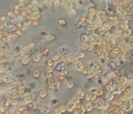
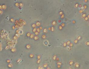
In the picture above, note the granulocytes among the red blood cells. Granulocytes are white blood cells that have small granules or particles. These contain proteins that help fight off viruses and bacteria. Three types of granulocytes are neutrophils, eosinophils, and basophils.
In the above picture, you can really see the shapes of the red blood cells, the depression in the middle of the round cell looks like a shadow. In the center is a white blood cell.
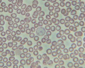
This specimen above was stained with Wright’s Stain, which stains the white blood cells blue. image was taken in brightfield lighting – no phase contrast, and you can see in the center a neutrophil white blood cell.

I hope this information was helpful. Please comment with your own experiences.